
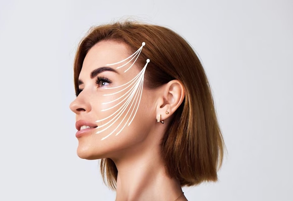
Prominent ear correction
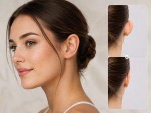
Prominent ear correction can be considered with Dr. Ömer Parıldar when the ears project noticeably away from the head or when differences in ear shape, position or symmetry create a persistent personal concern. The medical term commonly used for surgical reshaping of the external ear is otoplasty. Treatment does not aim to create identical ears or make them appear artificially close to the head. Instead, surgical planning focuses on understanding the cartilage features responsible for prominence and creating a position that remains proportionate to the head and face.
Prominent ears can be present from early life because of the natural way the cartilage develops. In some people, the fold in the upper ear is less defined. In others, the deeper bowl-shaped portion of the ear projects more strongly. These characteristics can occur individually or together.
One ear may also project more than the other.
Because the shape of each ear is different, the appropriate surgical technique can vary considerably between patients. Dr. Ömer Parıldar evaluates the cartilage structure, ear-to-head relationship, asymmetry and personal expectations before discussing a treatment plan.
The purpose of consultation is not simply to measure how far the ears project. It is to understand which structures create that projection and determine how much change would look natural for the individual patient.
Why Can Ears Appear Prominent?
The external ear is formed mainly from cartilage covered by relatively thin skin.
Its complex shape includes several folds and curves that determine how close the ear sits to the side of the head.
One important structure is the antihelical fold. When this natural fold is not strongly developed, the upper part of the ear can project outward.
Another factor is the concha, which is the deeper bowl-shaped part of the ear close to the ear canal. If this structure is particularly deep or prominent, the entire ear may sit further away from the head.
Some patients have both findings.
The earlobe can also contribute to overall prominence. Even after the upper cartilage is repositioned, a projecting earlobe may require separate consideration if it creates imbalance.
These anatomical differences are not caused by poor habits or something the patient has done incorrectly.
They are usually simply part of natural development.
Understanding which part of the ear creates the concern is important because different structures require different surgical techniques.
Prominent ear correction: Who May Consider It?
Prominent ear correction may be considered by patients who are bothered by the position or shape of their ears and who are medically suitable for surgery.
For adults, the decision is usually based on personal preference rather than medical necessity.
Some people have lived comfortably with prominent ears for many years and later decide that they would like to change them. Others have felt self-conscious about hairstyles, photographs or certain angles for a long period.
The reason for seeking treatment does not need to be dramatic.
A persistent personal concern can be enough to justify requesting information and discussing whether surgery is appropriate.
Children can also undergo ear reshaping in selected circumstances once the ears have reached sufficient development, but the timing and suitability require individual medical assessment and age-appropriate decision-making.
For every patient, realistic expectations are important.
Surgery can reduce projection and reshape selected cartilage features, but it cannot guarantee completely identical ears.
The two sides of the face and head are naturally asymmetric, and the ears usually have subtle differences even before treatment.
Dr. Ömer Parıldar evaluates these differences during consultation and explains which features may be improved and which natural variations may remain.
The Importance of Looking at Both Ears Separately
It is tempting to think of the ears as a perfectly matched pair.
In reality, most people have some degree of asymmetry.
One ear may sit slightly higher.
One may have a deeper conchal bowl.
The cartilage folds can also have different shapes.
Sometimes one ear is clearly more prominent than the other.
Treatment therefore should not automatically use exactly the same manoeuvre on both sides.
Dr. Ömer Parıldar assesses each ear independently before considering how they relate as a pair.
The aim is improved balance.
Trying to create absolute symmetry can lead to unnecessary overcorrection because the surrounding skull and facial structures are not perfectly symmetrical either.
Photographs taken from the front, sides and back may be useful for planning.
These views help show how much each ear projects and whether the position changes at different levels from the upper ear to the earlobe.
The patient should also explain which difference bothers them most.
Some people are concerned primarily about the upper ear.
Others notice the entire ear projecting outward.
The treatment can then be planned around the actual concern rather than around a general idea of making the ears smaller.
Does Ear Correction Make the Ears Smaller?
Otoplasty primarily changes shape and position.
It does not necessarily reduce the overall size of the ear.
A prominent ear may look visually smaller after it has been repositioned closer to the head, even if very little cartilage has been removed.
In some patients, selected cartilage may need to be reduced or reshaped.
However, removing tissue is not the main objective in every operation.
Patients who feel that their ears are physically too large should explain this during consultation because true ear-size reduction is different from simply reducing prominence.
The external ear has multiple dimensions.
Height, width, projection and earlobe size can all influence perception.
Dr. Ömer Parıldar evaluates which of these characteristics is responsible for the concern before discussing how surgery may change the appearance.
Prominent ear correction: How Is Surgery Planned?
Prominent ear correction begins with careful examination of the cartilage. Dr. Ömer Parıldar considers the antihelical fold, conchal depth, earlobe position, ear height and the relationship between the ear and the side of the head.
The skin behind the ear is also assessed.
Previous scars, injuries or ear operations should be discussed because they can influence surgical planning.
The patient’s desired degree of change matters as well.
Some people want a relatively subtle repositioning.
Others have significant projection and are looking for a more noticeable reduction.
The plan should avoid pulling the ears excessively close to the head.
Overcorrection can look unnatural and may create an ear that appears flattened.
The ear also needs to maintain its natural curves rather than becoming a simple smooth surface.
Measurements may be taken during assessment, but surgical planning is not based on one ideal angle that applies to everyone.
Head shape and ear anatomy differ between patients.
The intended result should therefore fit the individual’s proportions.
How Is the Ear Cartilage Reshaped?
Different cartilage techniques can be used depending on what creates the prominence.
When the antihelical fold is underdeveloped, surgical sutures can be placed to create or strengthen the fold.
These sutures help shape the cartilage into a more defined curve.
When the conchal bowl contributes strongly to projection, the cartilage can be repositioned closer to the head.
In selected cases, a small amount of cartilage may be removed.
Some techniques reshape cartilage through controlled scoring or weakening.
Others rely more heavily on permanent internal sutures.
The chosen method depends on cartilage thickness, flexibility and the type of correction required.
Adult ear cartilage can be firmer than cartilage in younger patients, which may influence the surgical approach.
The objective is to obtain stable repositioning while preserving a natural surface contour.
A well-planned procedure does not treat cartilage as though it were identical in every patient.
Dr. Ömer Parıldar chooses the technique according to the specific ear structure rather than applying one standard method.
Where Is the Incision?
The incision for traditional otoplasty is commonly positioned behind the ear.
This allows access to the cartilage while placing the resulting scar in the natural crease between the ear and the head.
The length of the incision depends on the procedure.
Because it is located behind the ear, the scar is generally less noticeable from normal viewing angles once healing progresses.
However, it remains a permanent surgical scar.
During the first weeks or months, the area can appear pink or firmer than the surrounding skin.
Scar maturation continues gradually.
Patients with a history of thick or raised scars should mention this during consultation.
The skin behind the ear can occasionally develop prominent scarring in susceptible individuals.
Dr. Ömer Parıldar can explain the anticipated incision and scar location before surgery.
Understanding this in advance gives patients a realistic idea of what the procedure involves.
What Happens During the Operation?
The appropriate anaesthetic approach depends on the patient’s age, treatment extent, medical history and surgical plan.
After the ear position has been marked, the incision is created according to the chosen technique.
The underlying cartilage is then accessed.
If a new antihelical fold needs to be created, sutures can be placed to reshape the cartilage.
If excessive conchal prominence is present, the cartilage can be repositioned or selectively reduced.
The surgeon repeatedly evaluates the ear position during the procedure.
This is important because the goal is not simply maximum flattening.
The upper, middle and lower portions of the ear should sit in a natural relationship with the side of the head.
After the cartilage has been positioned appropriately, the incision is closed.
Dressings are then placed to protect the ears and help maintain their position during early healing.
The exact procedure can differ between the two ears when their anatomy is different.
Why Natural Positioning Matters
An ear that is too close to the head can look just as unnatural as one that projects excessively.
Normal ears do not lie completely flat.
They have a natural angle and visible contours.
The upper ear, middle portion and earlobe also do not necessarily project by exactly the same amount.
Surgical planning therefore needs to preserve this three-dimensional structure.
Another concern is the shape of the front surface.
If cartilage is bent or tightened too aggressively, visible ridges or unnatural folds may develop.
The objective should be a gradual curve rather than a sharply created line.
Dr. Ömer Parıldar evaluates the ear from multiple angles during planning because a position that looks appropriate from the front also needs to appear natural from the side and back.
The patient’s hairstyle can influence how frequently the ears are visible, but treatment should remain anatomically appropriate regardless of whether they are usually covered.
Prominent ear correction: Recovery
Prominent ear correction recovery typically includes swelling, tenderness and temporary sensitivity around the ears. The degree of swelling varies, and the ears can initially look tighter or closer to the head than expected.
This early appearance is not necessarily the final position.
The tissues need time to relax and settle.
A protective head dressing may be used immediately after the operation.
Once the initial dressing is removed, a softer headband can sometimes be recommended for a period, particularly during sleep.
The exact schedule depends on the technique and individual healing.
Patients should follow the instructions given by Dr. Ömer Parıldar rather than adjusting dressings or headbands independently.
The ears may feel numb in some areas.
Temporary changes in sensation are possible because small nerves around the skin have been affected during surgery.
Sensation often improves gradually.
Patients should avoid unnecessary pressure on the ears while healing.
How Should You Sleep After Surgery?
Sleeping position is an important practical concern after ear surgery.
Direct pressure on a freshly operated ear can create discomfort and unnecessary stress on the healing tissues.
Patients may therefore be advised to sleep on their back during the early recovery period.
Extra pillows can help reduce accidental turning.
A protective headband may also be recommended at night after the initial dressing period.
Patients who normally sleep on their side should prepare for this temporary change before surgery.
Soft pillows alone do not make direct pressure appropriate unless the treating team has specifically allowed side sleeping.
The ears can remain sensitive for some time even after the most obvious swelling has improved.
It is useful to think about sleeping arrangements as part of surgical preparation rather than trying to improvise after returning home.
Hair Washing and Everyday Care
Hair washing requires care because incisions are generally located behind the ears.
Patients should follow specific instructions about when washing can resume.
Water, shampoo and friction around a fresh incision may need to be limited during the early healing period.
When washing is permitted, the area should be handled gently.
Hairbrushes, combs and hairdryers can accidentally catch or irritate the ear.
Glasses can also place pressure around the ear depending on their design.
Patients who wear glasses throughout the day should discuss this before surgery.
The position of headphone bands, helmets or tight hats should also be considered.
Do not use accessories that place pressure on healing cartilage until they have been approved.
These daily details can seem minor before surgery, but they have a meaningful role in protecting the ears during early recovery.
Returning to Work or School
The return to normal daily activity depends on the extent of surgery and how visible swelling or dressings remain.
Some patients feel physically well relatively quickly but prefer additional time away because the ears are still swollen.
Work that does not involve physical contact usually requires less restriction than employment where the ears could be struck.
Patients should also consider uniforms or safety equipment.
Helmets, tight head protection or communication headsets may not be suitable during early healing.
For school-age patients, playground activity and accidental contact require particular consideration.
The timing of return should therefore take into account more than discomfort alone.
Dr. Ömer Parıldar provides guidance based on the procedure and healing progress.
Normal concentration and light daily movement can return before activities that place the ears at risk.
Exercise and Sports
Light activity can generally return before strenuous training.
Patients should avoid exercise that substantially increases the risk of trauma to the ears during early healing.
Contact sports require particular caution.
Football, basketball, martial arts, wrestling and similar activities can expose the ear to direct impact.
Even a relatively minor blow could interfere with healing cartilage.
Swimming also requires the incisions to be sufficiently healed before entering water.
Exercise involving helmets or tight headgear needs separate consideration because pressure around the ears can be problematic.
Patients who regularly participate in sports should explain their activities during consultation.
The return schedule should reflect the specific sport rather than simply dividing activities into exercise and non-exercise.
Protecting the ears during the initial healing period is important for maintaining the surgical correction.
Possible Risks and Complications
Every surgical procedure carries potential risks.
Bleeding can occur and may lead to a hematoma, which is a collection of blood beneath the skin.
A rapidly swelling or increasingly painful ear after surgery requires prompt medical assessment.
Infection is another possible complication.
Because cartilage has a different blood supply from many other tissues, infections involving cartilage require appropriate medical attention.
Delayed wound healing can also occur.
Scars may become thicker or more raised than expected.
Some patients are naturally more prone to prominent scar formation.
Changes in skin sensation can occur around the ears.
Asymmetry can remain or develop.
One ear may settle differently from the other during healing.
There is also a possibility that some degree of prominence can recur over time if cartilage or sutures change.
Overcorrection is another potential issue.
An ear that is positioned too close to the head can appear unnatural.
Cartilage irregularities or visible contours may also occur.
The purpose of informed consent is to understand these possibilities before deciding whether surgery is appropriate.
When Should You Contact the Medical Team?
Patients should receive clear postoperative instructions.
Increasing pain that does not follow the expected recovery pattern should be reported.
Rapid swelling, particularly when one ear suddenly becomes much larger than the other, requires assessment.
Increasing redness, warmth, unusual discharge or fever can indicate infection.
A dressing that feels excessively tight should also be discussed rather than simply removed without guidance.
Changes in skin colour around the ear may require evaluation.
Patients should not wait for a scheduled appointment when symptoms are rapidly worsening.
When in doubt, contacting the surgical team is more appropriate than repeatedly adjusting the ear or dressing at home.
Following a structured aftercare plan allows problems to be identified early when they occur.
How Does the Result Develop?
The change in ear projection is visible early, but swelling affects the initial appearance.
The ears may seem more tightly positioned immediately after surgery than they will after healing.
As swelling decreases and tissues settle, the contour generally becomes softer.
Minor differences between the ears can be more noticeable during early recovery because swelling does not always resolve at exactly the same rate.
Scar maturation takes longer.
The area behind the ear may remain pink or firm for a period before gradually changing.
Patients should evaluate the result over time rather than during the first days.
The cartilage also needs time to stabilise in its new position.
For this reason, protecting the ears from trauma remains important even after the patient feels comfortable.
The goal of Prominent ear correction is a balanced ear position that fits naturally with the patient’s head and facial proportions rather than ears that appear surgically flattened.
Can Ear Prominence Return?
Surgical correction is designed to create a long-lasting change, but some degree of recurrence is possible.
Cartilage naturally has elasticity and can exert forces against its new position.
Internal sutures can also change over time.
The likelihood of recurrence depends on the original anatomy, cartilage strength, surgical technique and healing.
Minor differences that develop over time do not always require further treatment.
If more significant prominence returns, the ears can be reassessed after adequate healing.
Revision surgery should not be considered too early because swelling and tissue adaptation can create temporary changes that are still evolving.
Patients should attend planned follow-up appointments so that the position can be evaluated progressively.
Prominent ear correction in Istanbul
Patients considering Prominent ear correction in Istanbul with Dr. Ömer Parıldar can begin with an individual assessment of cartilage shape, projection and symmetry.
If you are travelling from another city or country, mention this when scheduling the consultation.
The treatment plan should include sufficient time for early follow-up.
Patients may initially be wearing a protective dressing or headband, which should be considered when organising travel.
Avoid situations where the ears could be accidentally struck.
Heavy luggage is less directly related to the surgical area than after many body procedures, but crowded transport can still expose the ears to accidental contact.
Before returning home, understand how wound care should continue and whether sutures require removal.
You should also know when photographs or an in-person review may be requested.
A well-organised aftercare process is an important part of surgical treatment.
What Determines the Cost?
The cost depends on the individual surgical plan.
A patient requiring relatively simple cartilage folding may have a different procedure from someone who needs treatment of both the antihelical fold and conchal cartilage.
One-sided correction can also differ from treatment of both ears.
Anaesthesia and surgical facility requirements influence the overall plan.
Medical assessment and postoperative follow-up may also form part of the treatment structure.
For this reason, accurate pricing should follow examination.
Ask which elements of the procedure are included in the proposed estimate.
The decision about surgical technique should be based on the ear anatomy rather than on selecting the shortest or least expensive procedure.
A clear clinical plan makes the financial discussion much more meaningful.
Questions to Ask During Consultation
Ask which anatomical feature is making your ears appear prominent.
Find out whether the antihelical fold, conchal cartilage or earlobe contributes most.
Ask whether both ears require the same correction.
Discuss how much closer to the head the ears are expected to sit.
Ask where the incision will be placed.
Find out whether cartilage will be reshaped, repositioned or selectively reduced.
Ask how long a dressing or headband may be required.
Discuss when you can sleep on your side again.
If you play contact sports or regularly wear a helmet, mention this.
Ask when these activities can safely resume.
If one ear is naturally different from the other, ask how much asymmetry is expected to remain.
A useful consultation should provide a clear picture of the intended change while also explaining the limits of absolute symmetry.
Dr. Ömer Parıldar
For patients considering ear reshaping, Dr. Ömer Parıldar evaluates the cartilage folds, conchal structure, earlobe position, ear projection and natural differences between the two sides before discussing treatment.
You do not need to arrive knowing which otoplasty technique you require.
Begin by explaining which part of the ear concerns you and how much change you would like to see.
Some patients want only a subtle reduction in projection, while others have more pronounced prominence that requires broader cartilage reshaping.
An individual consultation allows the surgical technique, expected scar, healing process and realistic degree of correction to be discussed clearly.
The objective is not to eliminate the natural shape of the ear but to create a more balanced relationship between the ears, head and face.
Contact Dr. Ömer Parıldar
Telephone: +90 546 198 81 35
WhatsApp: +90 546 198 81 35
Email: info@dromerparildar.com
Address:
Basınköy, Florya Cd. No:15-17
34153 Bakırköy / İstanbul
Contact Dr. Ömer Parıldar to arrange an individual consultation and discuss a carefully planned approach to ear positioning and reshaping based on your cartilage structure, symmetry and personal expectations.
İlgili bağlantı: https://www.dromerparildar.com/iletisim/
İlgili bağlantı: https://www.instagram.com/dromerparildar/
Articoli correlati
Scopri altri approfondimenti dedicati ad argomenti simili.